
P7D
Delayed implants at 4.6 and 4.7 with laminate bone grafting
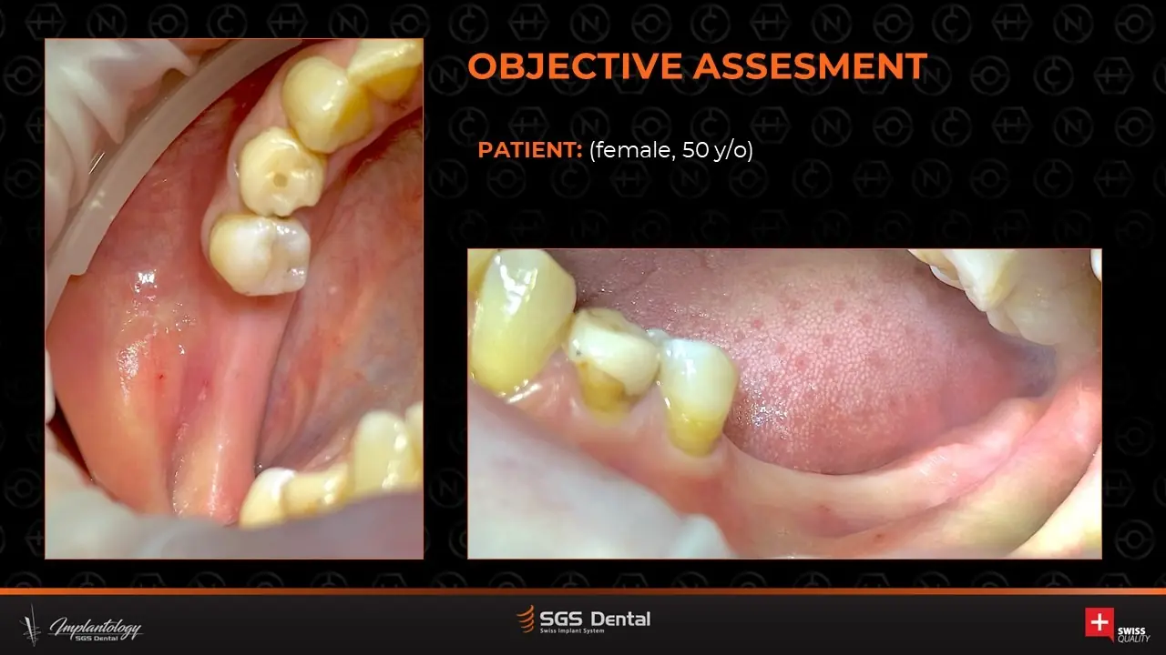
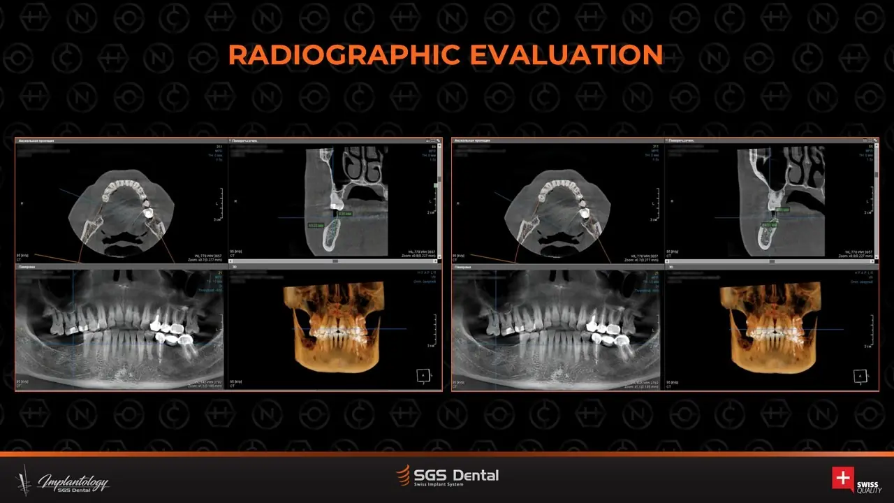
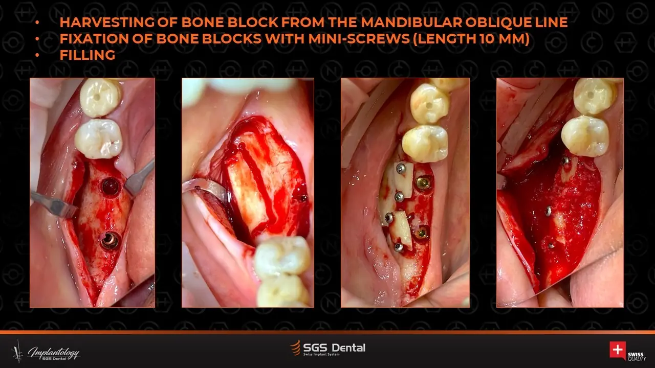
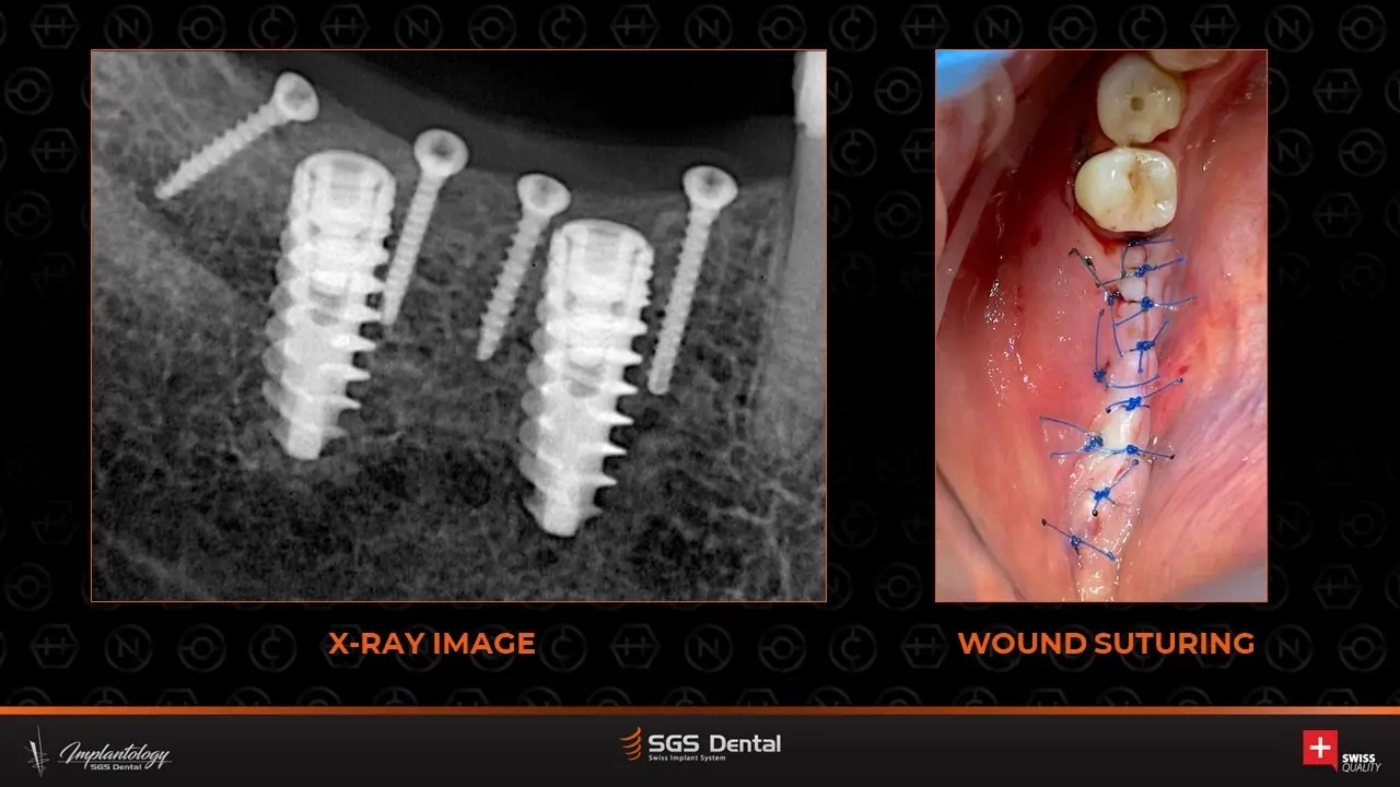
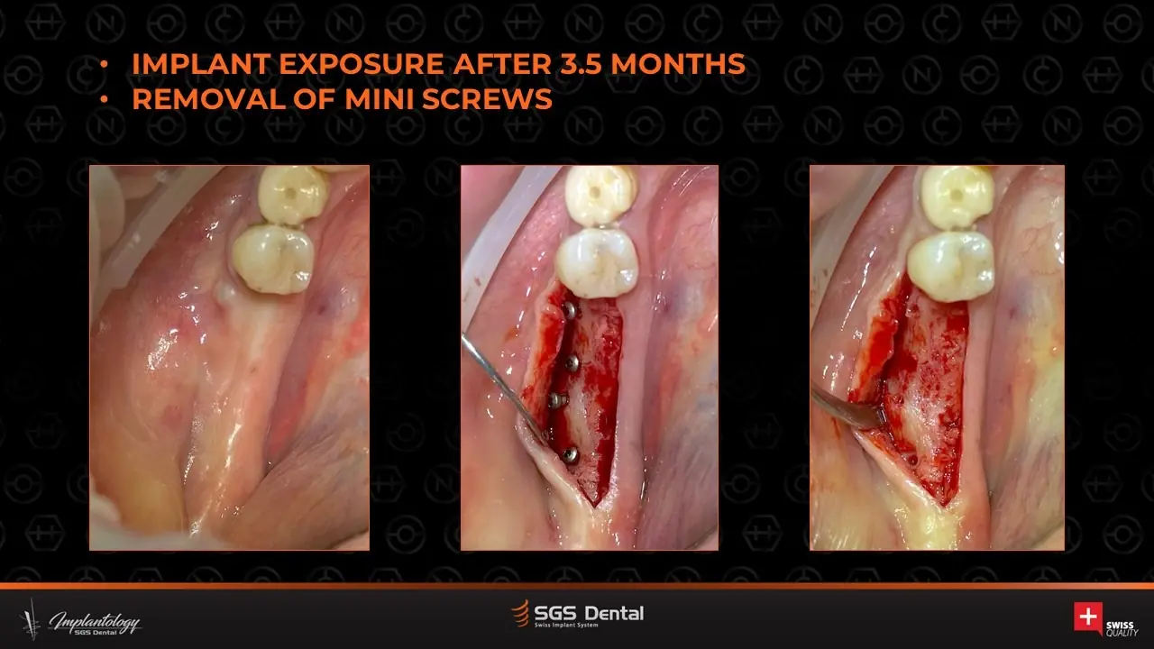
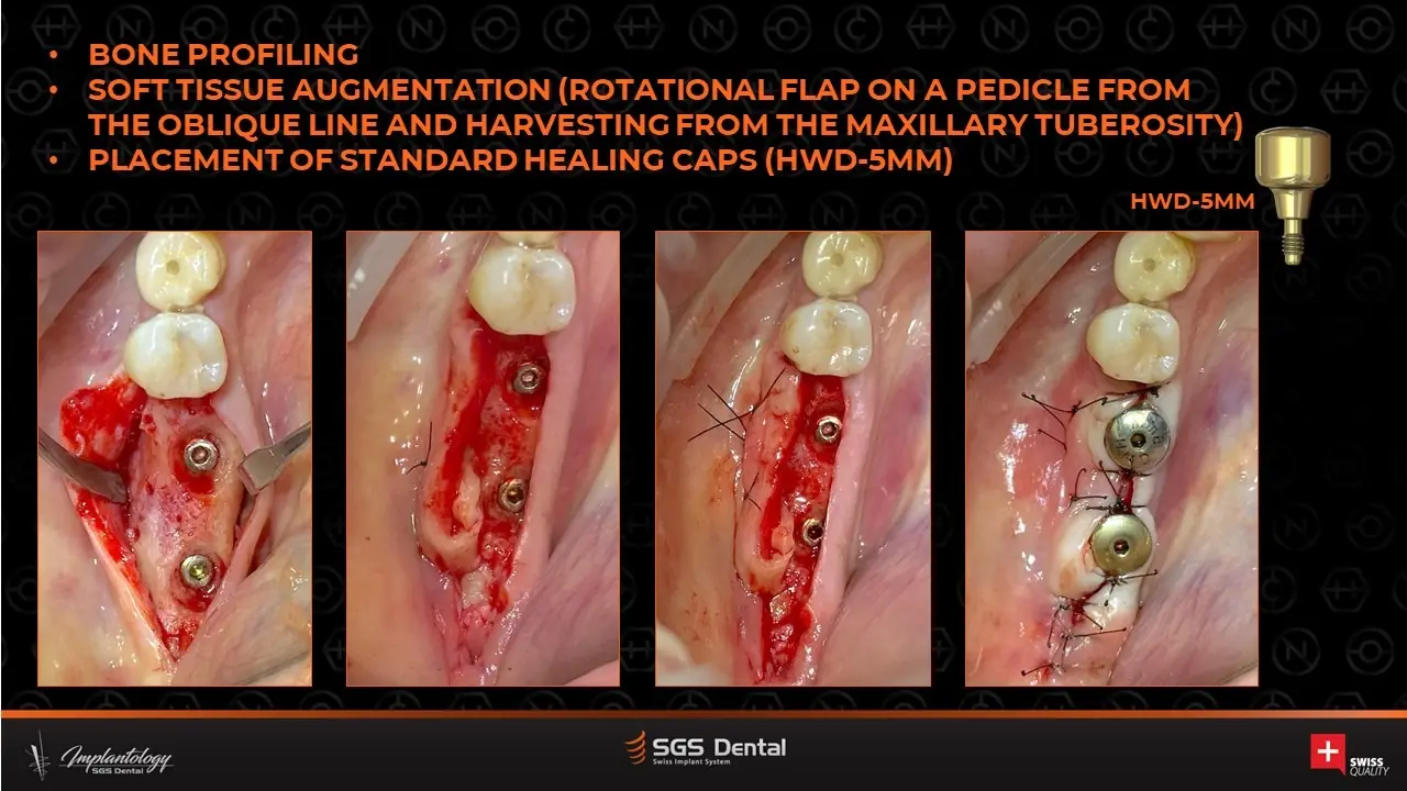
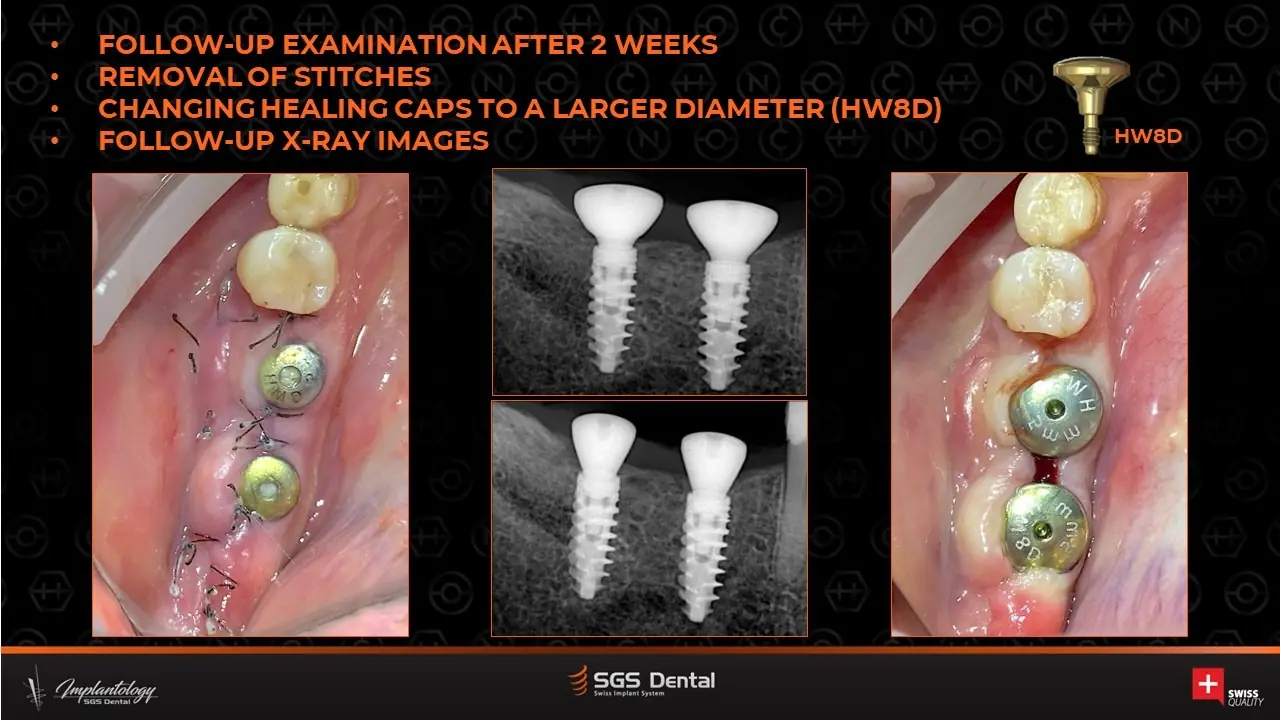
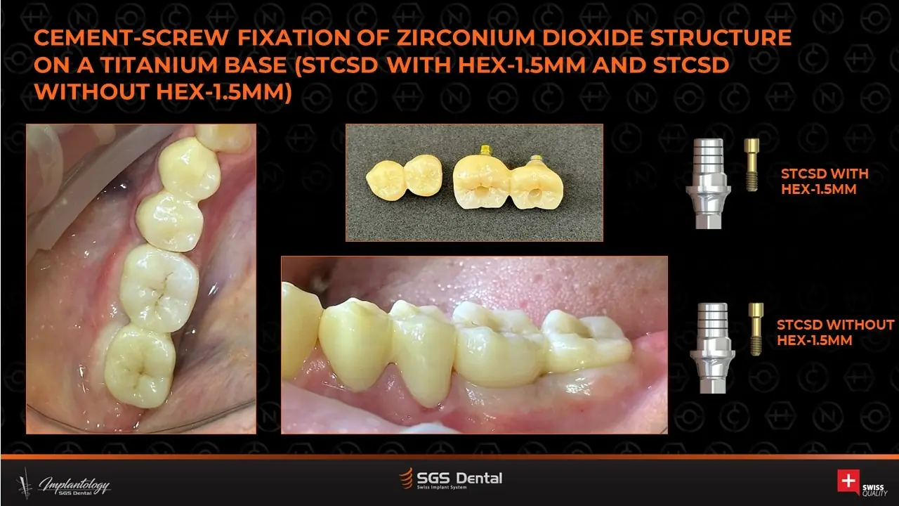
A 50-year-old woman. P7D implants were placed and the deficient ridge rebuilt with a bone block from the external oblique line fixed with mini-screws (laminate technique). At 3.5 months the implants were exposed, screws removed, bone profiled and soft tissue augmented; final zirconia on titanium bases was fixed with a cement-screw technique.
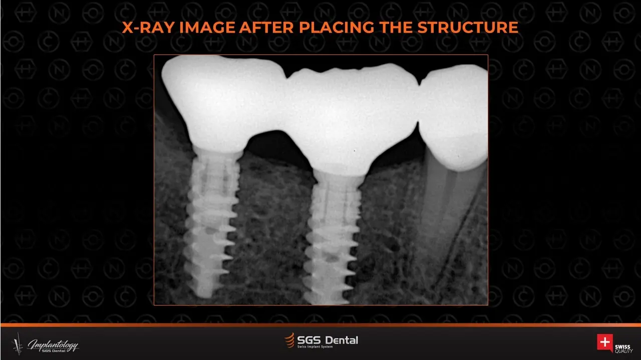
Timeline
Step by step
- Assessment and radiographs
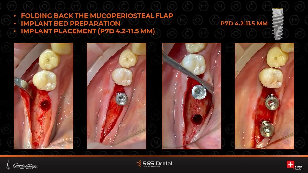
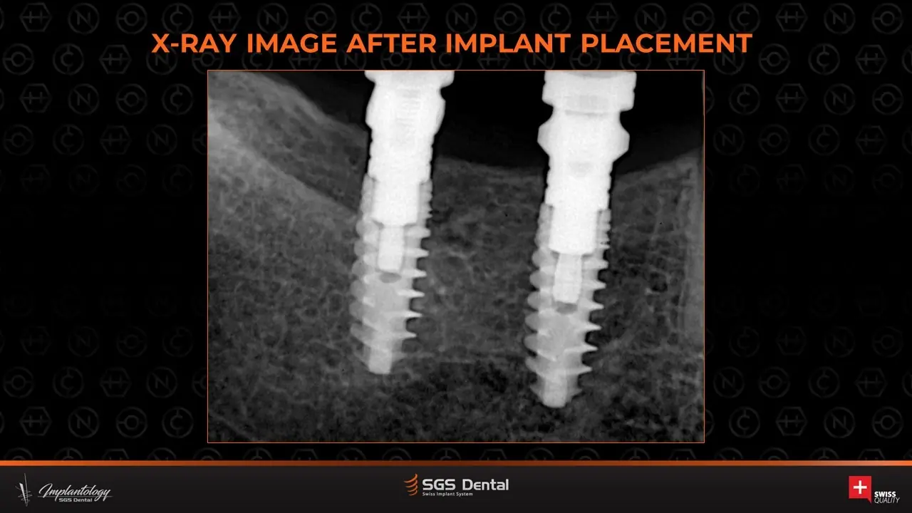
- Flap; P7D 4.2×11.5 mm placed
- Bone block from the mandibular oblique line fixed with 10 mm mini-screws; filling
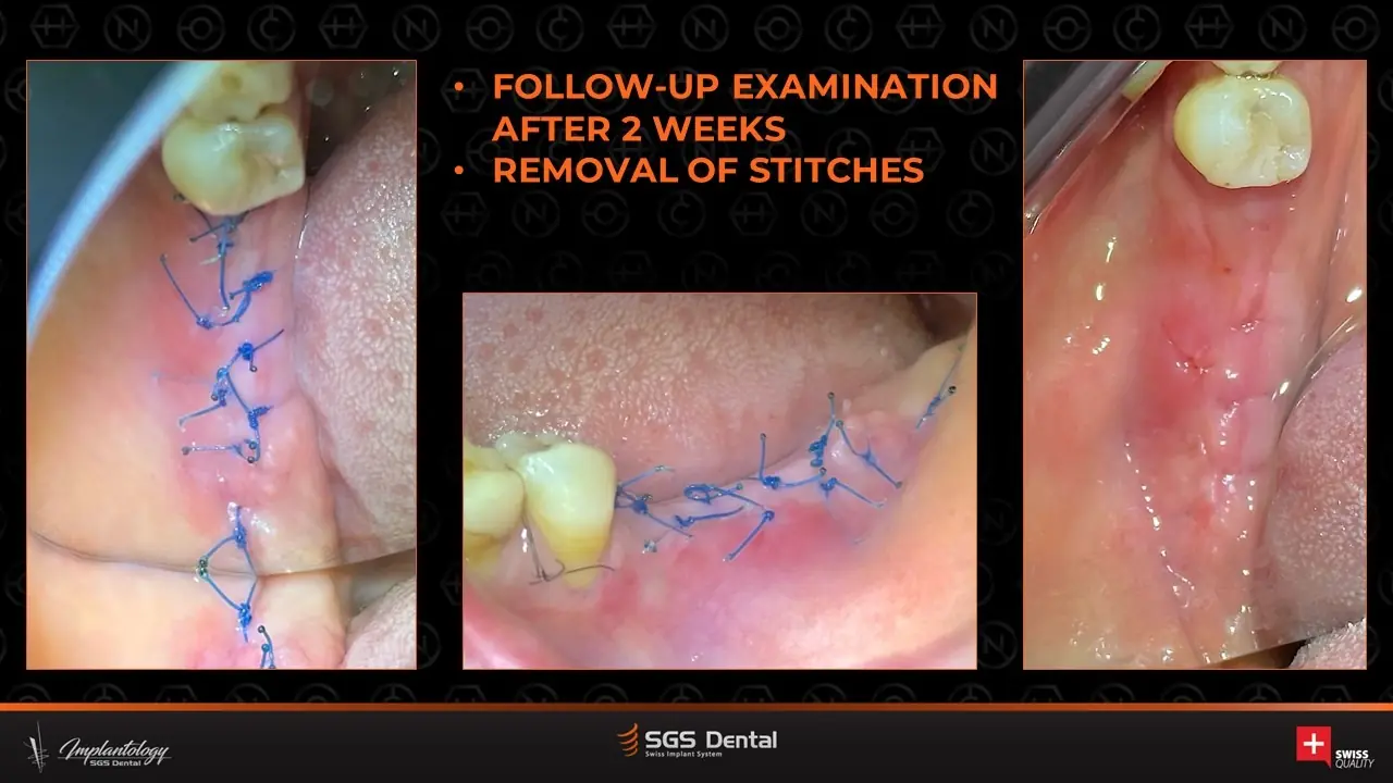
- 2 weekssuture removal
- 3.5 monthsexposure; mini-screws removed
- Bone profiling; soft-tissue augmentation (rotational pedicle flap + tuberosity graft); HWD-5 mm caps
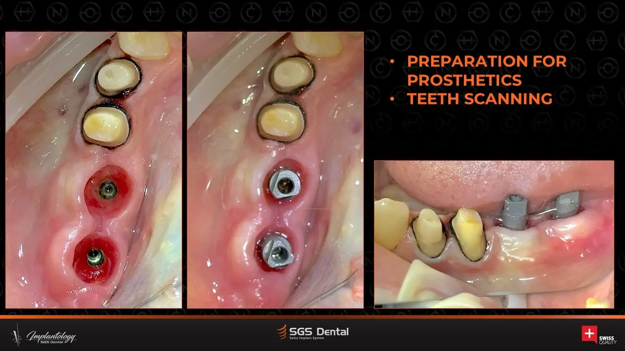
- 2 weekswider HW8D caps; scanning
- Cement-screw zirconia on STCSD 1.5 mm titanium bases
More cases
Similar cases
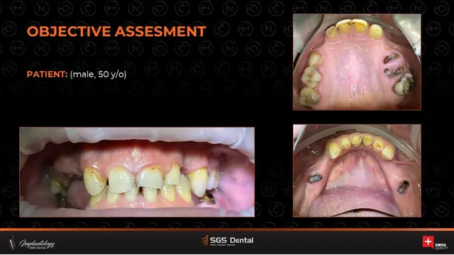
P5DP7D
Total rehabilitation of the dentition and jaw system
Full mouth – multiple segments, standard and single-stage (immediate) placement
View case →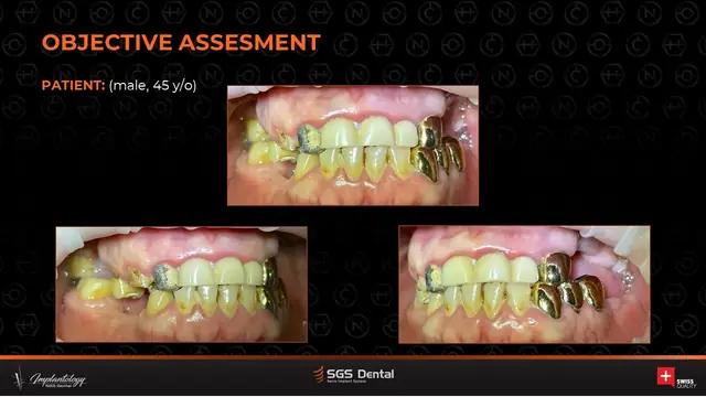
P5GP7D
Total rehabilitation of the dental system with P5G and P7D
Full mouth – posterior implants in both jaws with tooth-borne restorations
View case →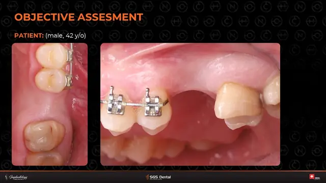
P7D
Open sinus lift with implant placement at tooth 2.6
Posterior maxilla – lateral-window sinus lift with simultaneous implant
View case →Request a quote / trial kit
Pricing, tailored to your practice.
Planning a similar case? Tell us about it and we'll send a private quote or a trial kit for the implants it needs.