
P5G
Immediate implant at 4.6 with bone cores
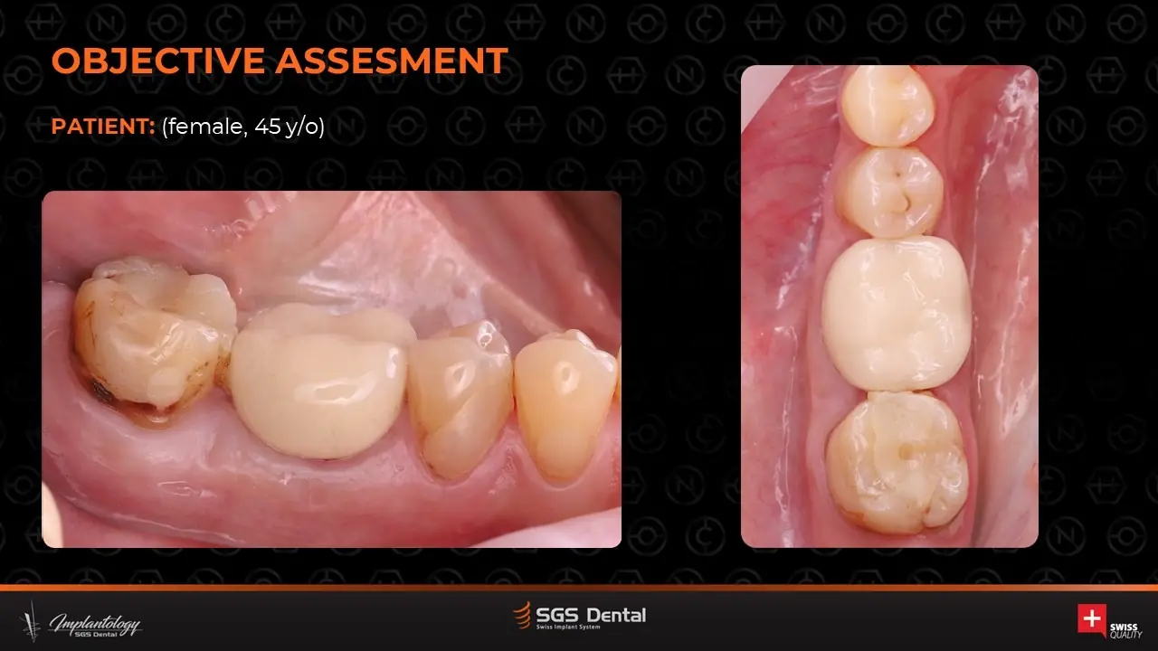
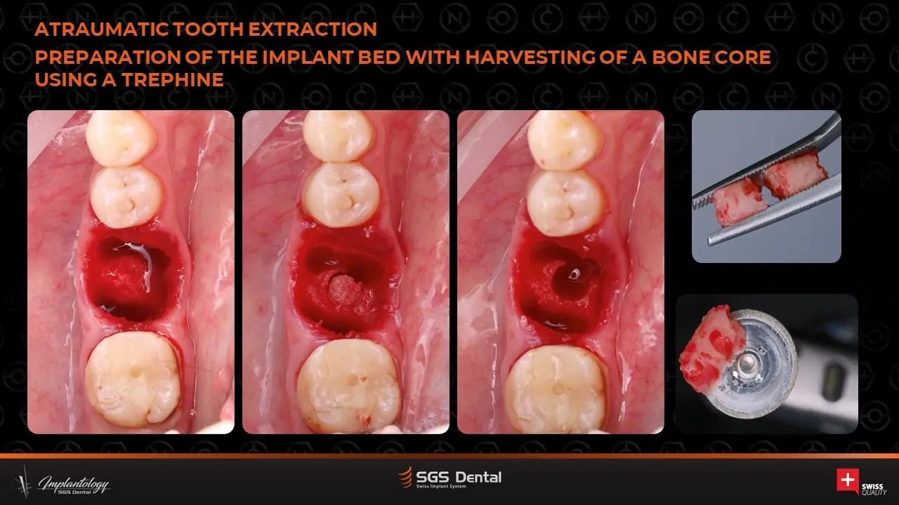
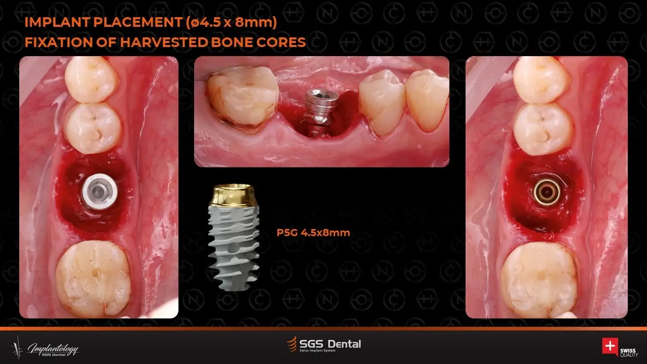
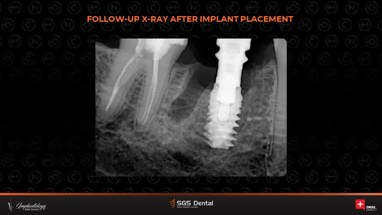
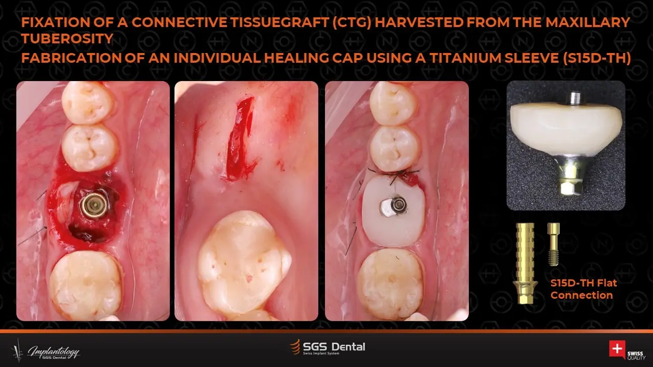
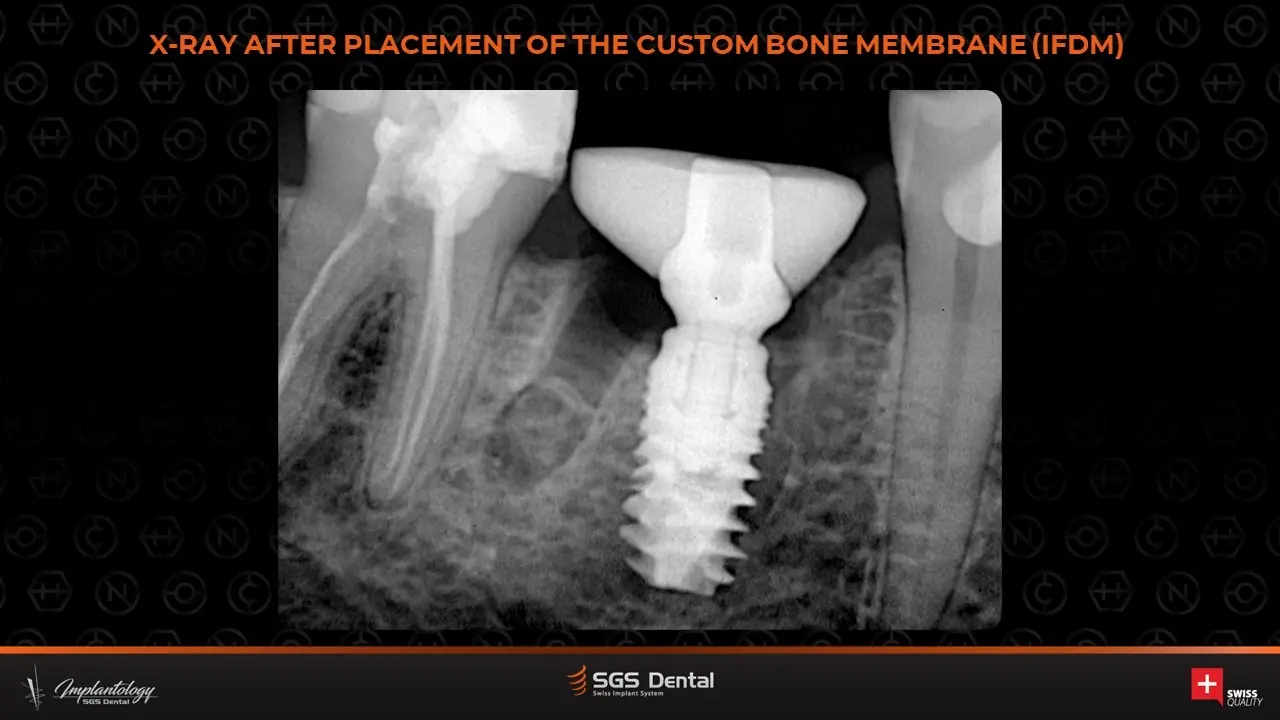
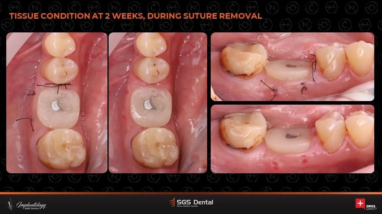
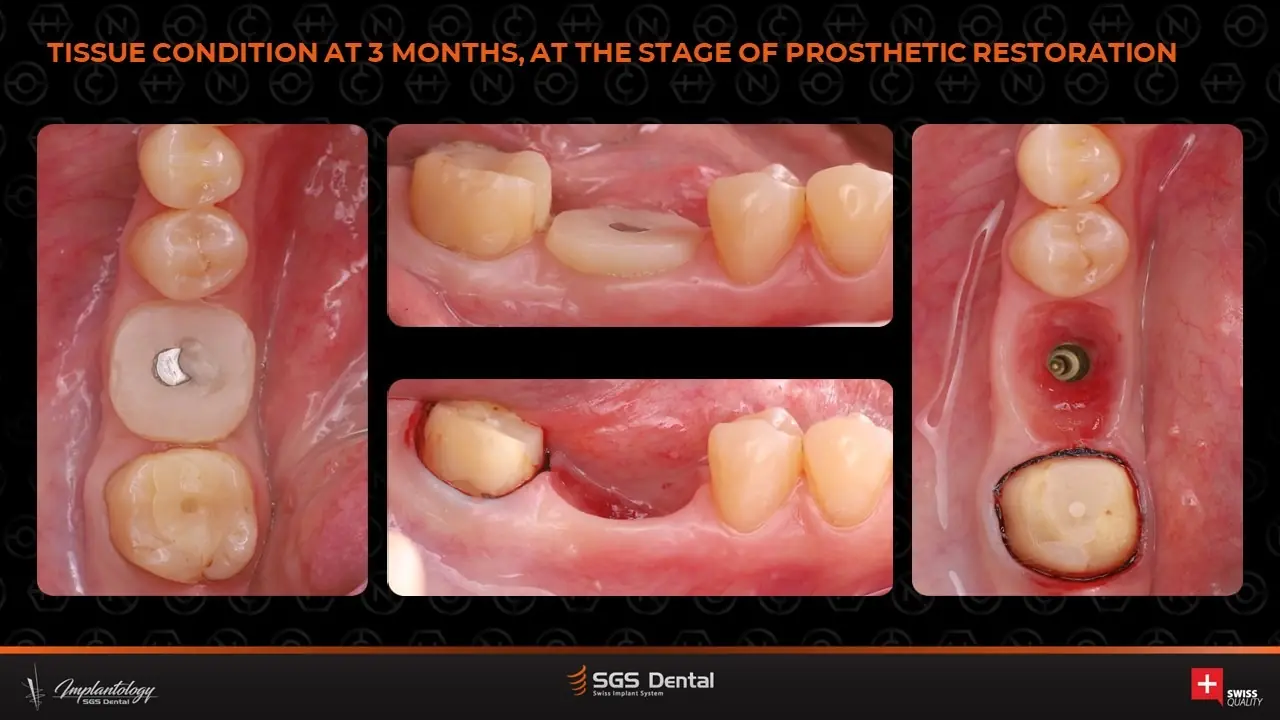
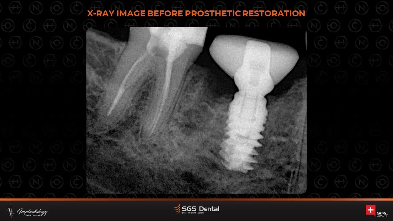
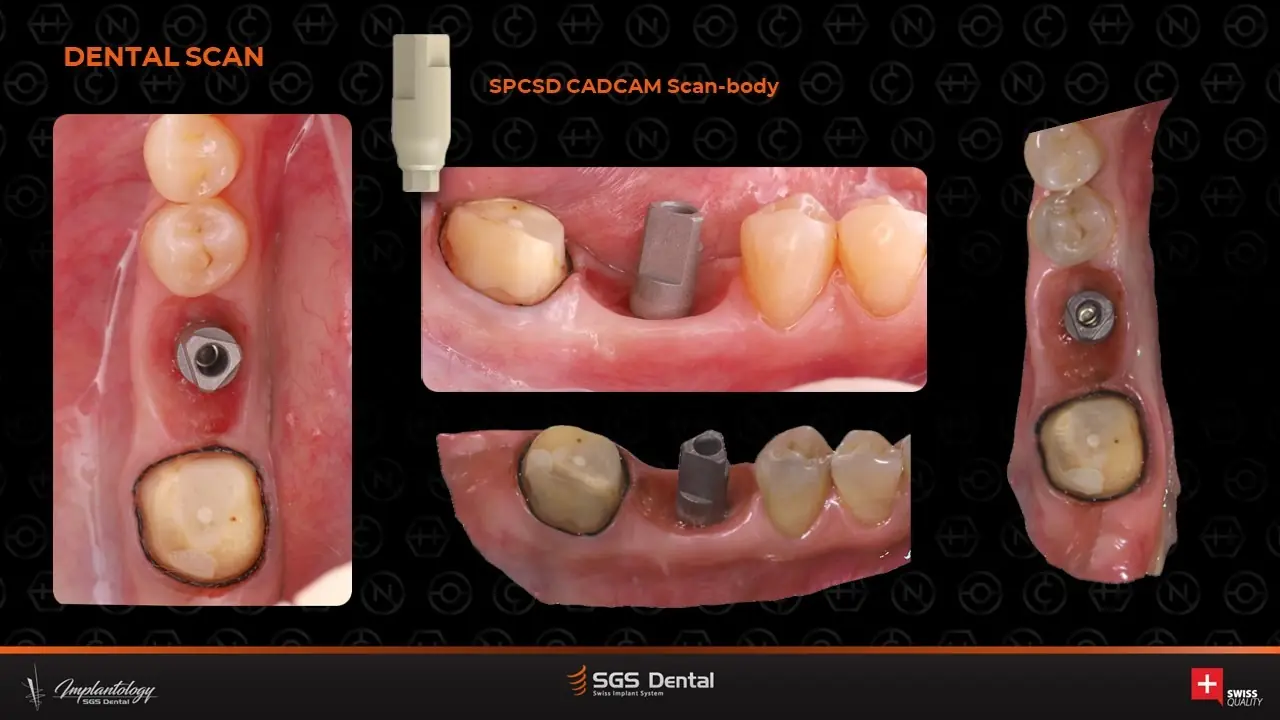
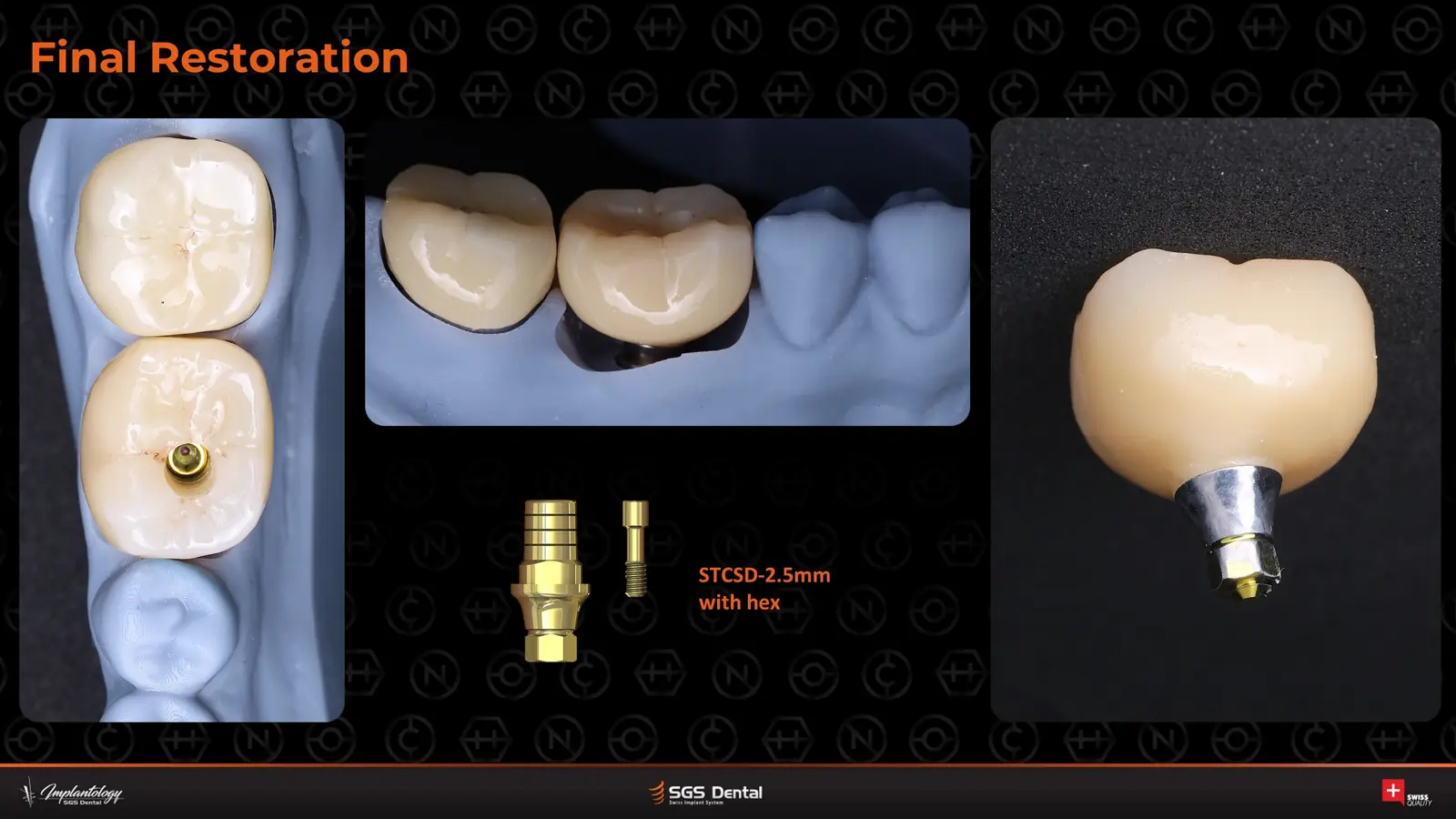
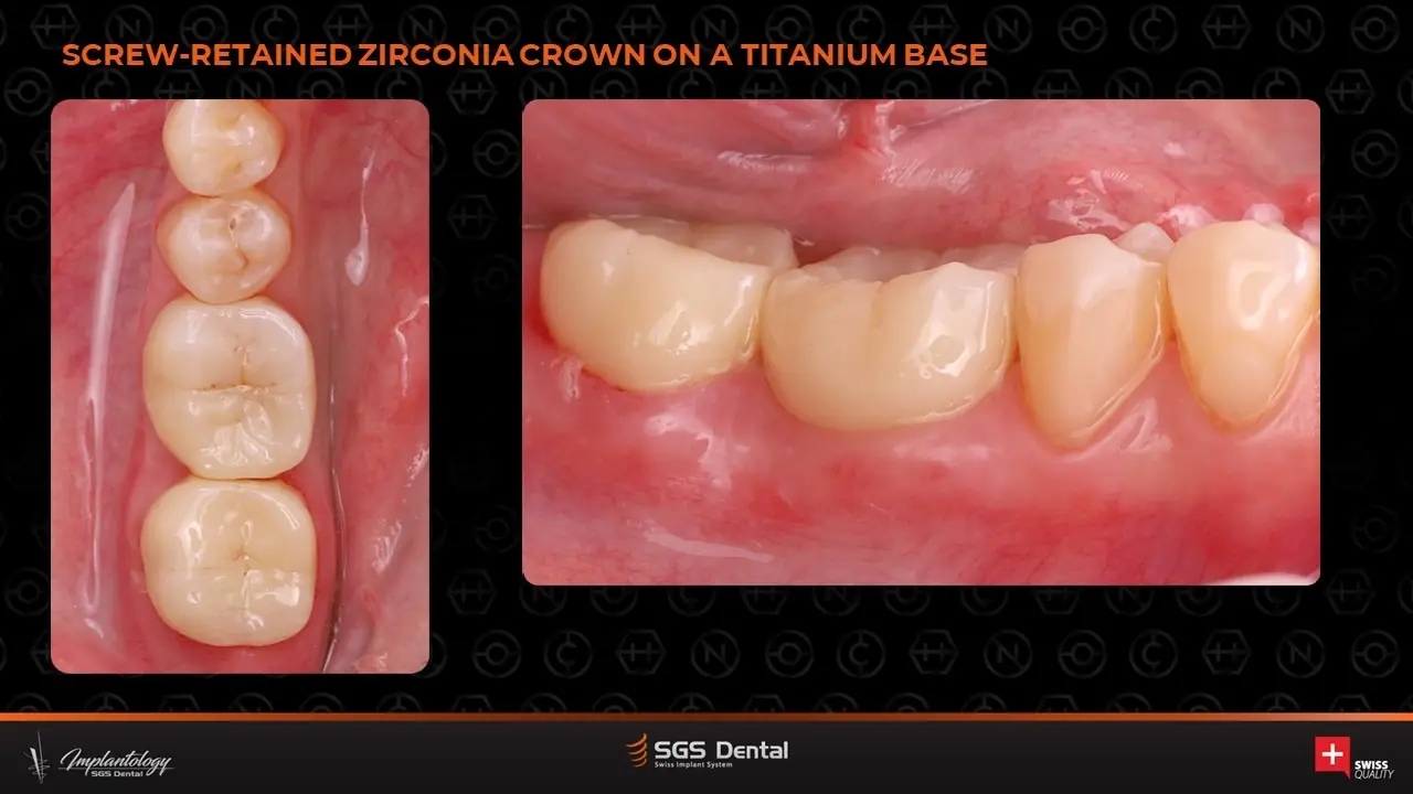
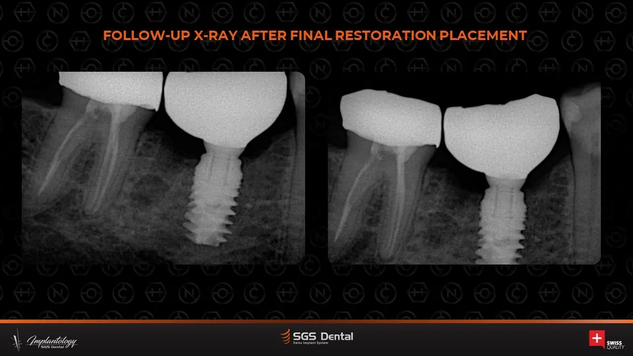
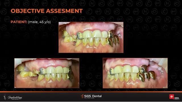
A 45-year-old woman. The molar was extracted, the bed prepared with a trephine and the harvested bone cores fixed around a P5G implant. A tuberosity CTG and a custom healing cap on an S15D-TH sleeve shaped the tissue. At 3 months the site was scanned with an SPCSD scan body and a screw-retained zirconia crown on a titanium base delivered.
Timeline
Step by step
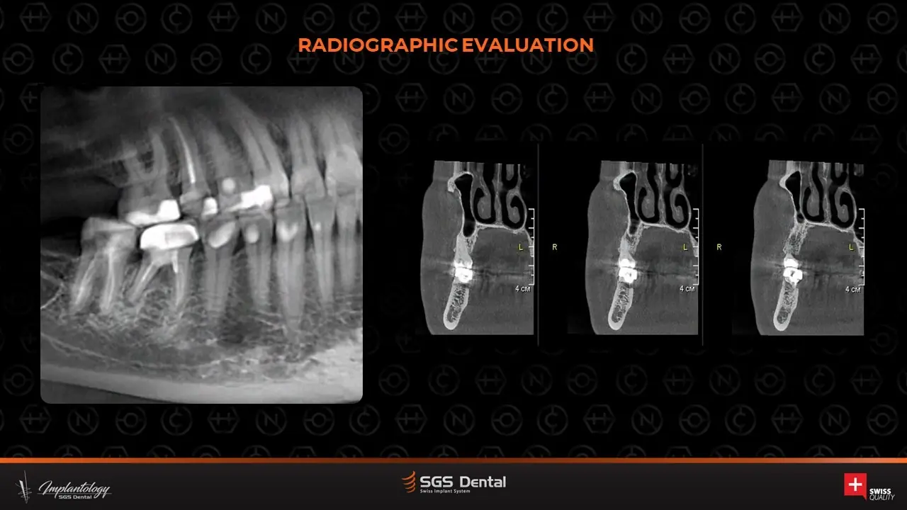
- Assessment and radiographs
- Atraumatic extraction; implant bed with trephine bone-core harvest
- P5G 4.5×8 mm placed; bone cores fixed
- Tuberosity CTG; custom healing cap on S15D-TH
- 3 monthstissue check and X-ray
- Digital scan with SPCSD scan body
- Screw-retained zirconia crown on titanium base (STCSD 2.5 mm with hex)
More cases
Similar cases
P5GP7D
Total rehabilitation of the dental system with P5G and P7D
Full mouth – posterior implants in both jaws with tooth-borne restorations
View case →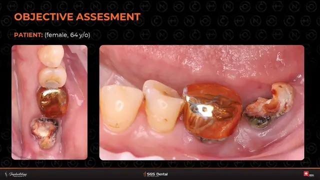
P5G
Immediate implants at 3.6 and 3.7 with retromolar bone graft
Molars – immediate implants with cyst removal and bone graft
View case →
P5GP1D
Navigated (fully guided) implantation in the upper jaw
Guided surgery – partial edentulism, upper jaw
View case →Request a quote / trial kit
Pricing, tailored to your practice.
Planning a similar case? Tell us about it and we'll send a private quote or a trial kit for the implants it needs.