The four graft families, in one paragraph each
Autograft. The patient's own bone, from the osteotomy, a trephine core or the ramus. It is the only option that brings living bone-forming cells, which is why it is called osteogenic. The trade-offs are limited volume and, for block harvests, a second surgical site. See a trephine core reused as graft in our bone core case at 4.6.
Allograft. Processed human donor bone, mineralized or demineralized. It is osteoconductive, and in socket studies it left the least residual graft at re-entry, which means it remodels into native bone relatively quickly (De Risi 2015).
Xenograft. Usually deproteinized bovine or porcine bone. It is osteoconductive and resorbs slowly. That slow substitution is exactly why the ITI consensus recommends low-substitution-rate materials and membranes when ridge preservation is followed by late implant placement (ITI).
Alloplast. Synthetic calcium phosphates and related materials. They are consistent and donor-free. In one large review of ridge augmentation they had the smallest average horizontal gain, about 2.2 mm, though the difference from other materials was not statistically significant (Troeltzsch 2016).
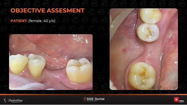
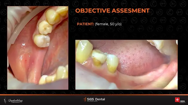
Extraction sockets
Unassisted healing is not neutral. Re-entry studies show 29 to 63% loss of ridge width and 11 to 22% loss of height in the first 6 months, with most of the change in the first 3 to 6 months (Tan 2012).
Socket grafting reduces that loss. In a meta-analysis of 22 randomized trials, ridge preservation saved about 2 mm of width compared with extraction alone, and particulate xenograft or allograft covered with a collagen membrane or a rapidly absorbable collagen sponge gave the most favorable horizontal results. Sites with a buccal wall thicker than 1 mm did better still (Avila-Ortiz 2019).
How long to wait? A histology meta-analysis found no major differences between graft types, and concluded that in preserved sites there is little reason to wait more than 3 to 4 months before placing the implant (De Risi 2015). If the plan is to place early, ITI describes type 2 placement after soft tissue healing, typically 4 to 8 weeks after extraction (ITI).
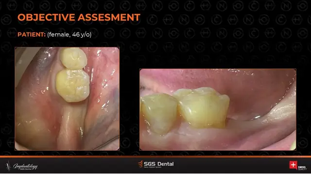
Horizontal ridge defects
For lateral augmentation, guided bone regeneration with particulate graft gained about 3.6 mm at the time of regeneration across randomized trials. About 1.2 mm of that resorbed within 6 months, and the authors recommend overcorrecting the contour to compensate (Elnayef 2018). A resorbable collagen membrane over a particulate graft remains the workhorse for contained, mostly horizontal defects.
See it in practice: the autogenous chip technique over a thin buccal wall in our 4.7 case, and a laminate bone block in the 4.6 and 4.7 case.
Vertical ridge defects
Vertical gain is the hardest thing we ask of a graft. A meta-analysis of 36 publications found that GBR averaged 4.18 mm of vertical gain with a 12.1% complication rate, compared with 3.46 mm and 23.9% for bone blocks. Within GBR, non-resorbable, space-making membranes reached about 4.4 mm. In the studies that reported it, re-entry took place between 4.6 and 10.4 months (Urban 2019). Plan for space maintenance, tension-free closure and a longer wait.
Sinus augmentation
For the lateral window, the graft matters less than many of us were taught. Implant survival after sinus floor augmentation appears to be independent of the biomaterial used (Al-Nawas 2014). Autograft still forms the most new bone histologically, but bone substitutes are a valid alternative that spares the patient a donor site. Biopsies taken after more than 4.5 months of healing showed significantly more new bone than earlier ones (Danesh-Sani 2017).
Two examples from our cases: a lateral-window lift with a simultaneous implant at 2.6 and a crestal lift using the Fugazzotto technique.
A quick decision table
| Defect | Common first choice | Membrane | Typical wait before placement or re-entry |
|---|---|---|---|
| Extraction socket | Particulate xenograft or allograft | Collagen membrane or sponge | About 3 to 4 months (De Risi 2015) |
| Horizontal | Particulate GBR, often autograft mixed with a substitute; overbuild | Resorbable collagen | Varies by protocol; expect about 1.2 mm resorption by 6 months (Elnayef 2018) |
| Vertical | GBR with space maintenance | Non-resorbable or reinforced | 4.6 to 10.4 months in the reviewed studies (Urban 2019) |
| Sinus (lateral) | Autograft, substitute or a mix | Per operator protocol | More new bone after 4.5 months (Danesh-Sani 2017) |
Rule of thumb. Autograft is the only osteogenic option. Allograft remodels faster. Xenograft resorbs slowly and holds contour.
Get each new clinical guide by email.
Twice a month, one short, referenced guide for implant dentists and surgeons: what the current evidence says, linked to real cases. Unsubscribe anytime.
- Summaries of current systematic reviews
- Surgical and restorative topics
- Real cases and practical protocols
Educational content for licensed dental professionals. It summarizes published reviews and is not a substitute for clinical judgment.
Sources
- Tan WL et al. Clin Oral Implants Res. 2012;23 Suppl 5:1-21. doi.org
- Avila-Ortiz G, Chambrone L, Vignoletti F. J Clin Periodontol. 2019. doi.org
- De Risi V et al. Clin Oral Implants Res. 2015. doi.org
- Elnayef B et al. Int J Oral Maxillofac Implants. 2018. doi.org
- Urban IA et al. J Clin Periodontol. 2019;46 Suppl 21:319-339. doi.org
- Al-Nawas B, Schiegnitz E. Eur J Oral Implantol. 2014. pubmed.ncbi.nlm.nih.gov
- Danesh-Sani SA et al. J Periodontal Res. 2017. doi.org
- Troeltzsch M et al. J Craniomaxillofac Surg. 2016. doi.org
- ITI Consensus: Implants in Postextraction Sites. academy.iti.org